Figure 1.
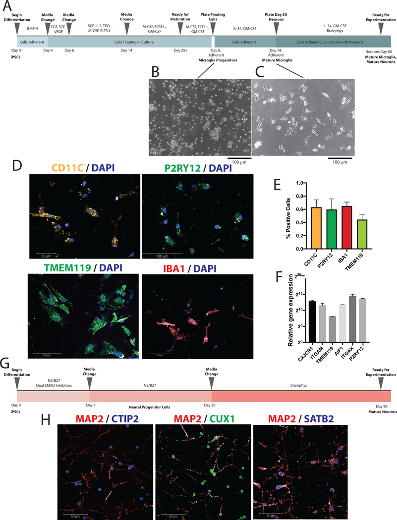
Differentiation and validation of iPSC-derived microglia. A. Schematic depiction of microglial differentiation from iPSCs through microglial maturity and co-culture with cortical neurons. B. Representative image of microglial progenitor cells after re-plating following day 25 of differentiation at 10x magnification. C. Representative image of mature microglia at day 14 at 10x magnification. D. Immunocytochemistry staining of CD11c, P2RY12, IBA1, and TMEM119 to confirm expression of microglial markers, shown at 20x magnification. E. Percentage of cells positively stained for microglial markers CD11c, P2RY12, IBA1, and TMEM119. F. qPCR validation showing microglia exhibiting microglial-signature genes including AIF1, CX3CR1, ITGAM, ITGAX, P2RY12, and TMEM119. G. Schematic depicting cortical neuron differentiation from iPSCs. H. Immunocytochemistry staining of CTIP2, CUX1, SATB2, and MAP2 to confirm generation of cortical neurons, shown at 63X magnification.

