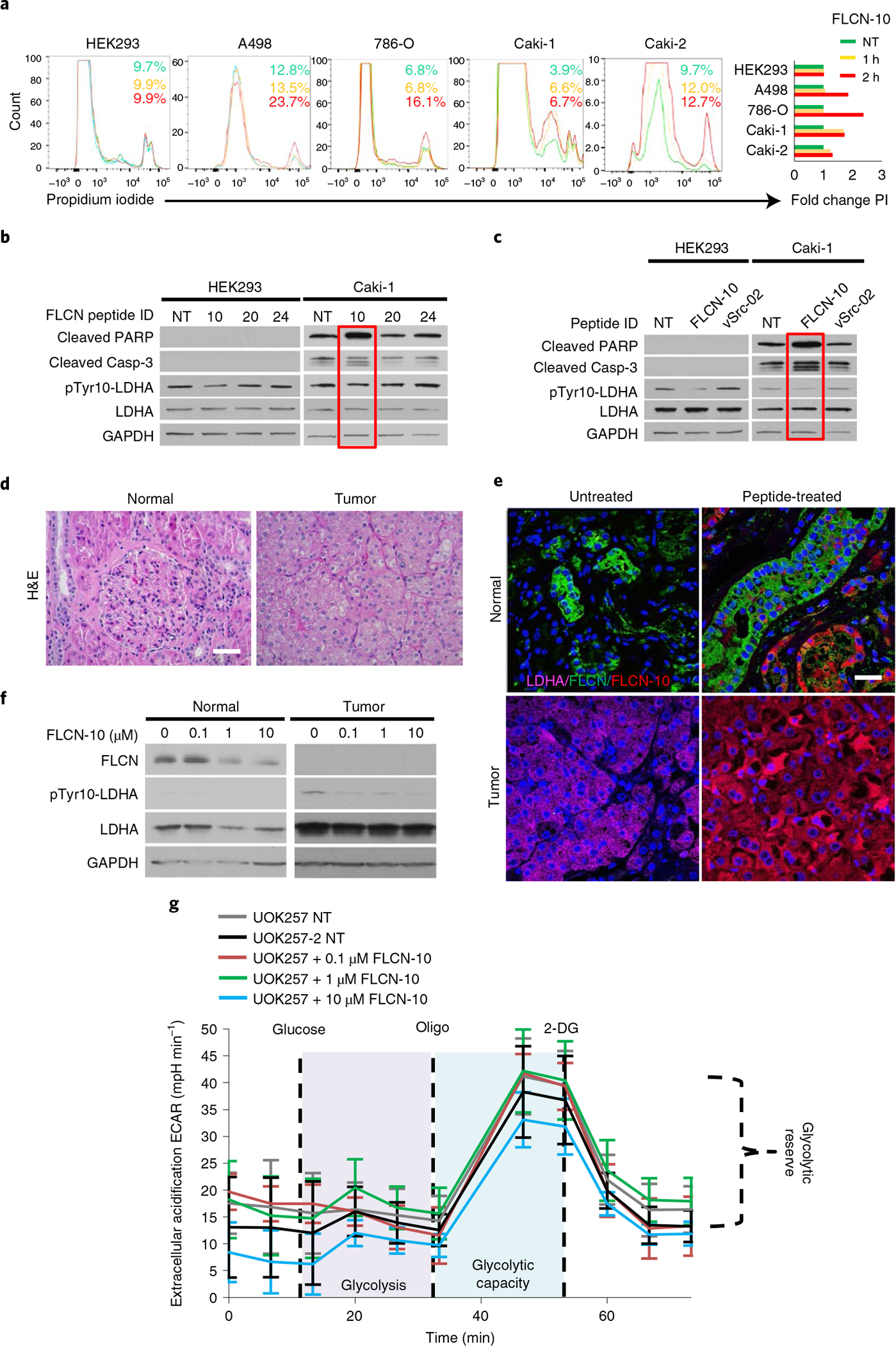
Fig. 5 |. FLCN peptide inhibits LDHA and induces apoptosis in cancer cells.

a, Flow-cytometric assessment of cell death in renal cell lines following treatment with 1 μM FLCN-10 for 1 h (yellow) or 2 h (red), as determined by propidium iodide staining. Green indicates no treatment (NT). A representative of three independent experiments is shown. b, Immunoblot of whole-cell lysates from HEK293 and Caki-1 cells following 1 μM treatment with the indicated FLCN peptides for 2 h. The blots are a representative example of three independent experiments. c, Immunoblot of whole-cell lysates from HEK293 and Caki-1 cells following 1 μM treatment with either FLCN-10 or vSrc-02 peptide for 2 h. d, Hematoxylin and eosin (H&E) staining of normal and tumor renal tissues from a patient with BHD. Scale bar, 40 μm. e, Fluorescence microscopy of normal and tumor renal tissues from a patient with BHD treated ex vivo with FLCN-10–rhodamine B and stained with anti-FLCN (green), anti-LDHA (pink) and DNA (Hoechst 33258; blue) Scale bar, 20 μm. f, Western blots of whole-cell lysates collected from e. g, Extracellular acidification rate of UOK257 cells treated with increasing amounts of FLCN-10 peptide for 2 h. Data are presented as mean ± s.d. (n = 4 independent samples). Source data for b, c, f and g are available online.
