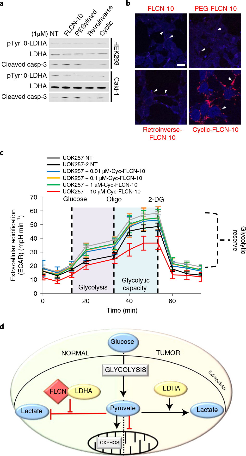
Fig. 6 |. Model of FLCN-mediated regulation of LDHA.

a, Western blot of pTyr10-LDHA and cleaved caspase-3 in HEK293 and Caki-1 cells following treatment with modified FLCN peptides. NT, no treatment. The blots are a representative example of three independent experiments. b, Fluorescence imaging of modified rhodamine B-labeled FLCN-10 peptides in HEK293 cells. White arrowheads denote examples of FLCN-10 localization. Scale bar, 10 μm. c, Extracellular acidification rate of UOK257 cells treated with increasing doses of cyclic-FLCN-10 peptide for 2 h. Data are presented as mean ± s.d. n = 6 independent samples. d, In normal cells, FLCN regulates the activity of LDHA. In cancer cells experiencing the Warburg effect, FLCN and LDHA have lost the ability to interact, leading to the hyperactivity of LDHA. Source data for a and c are available online.
