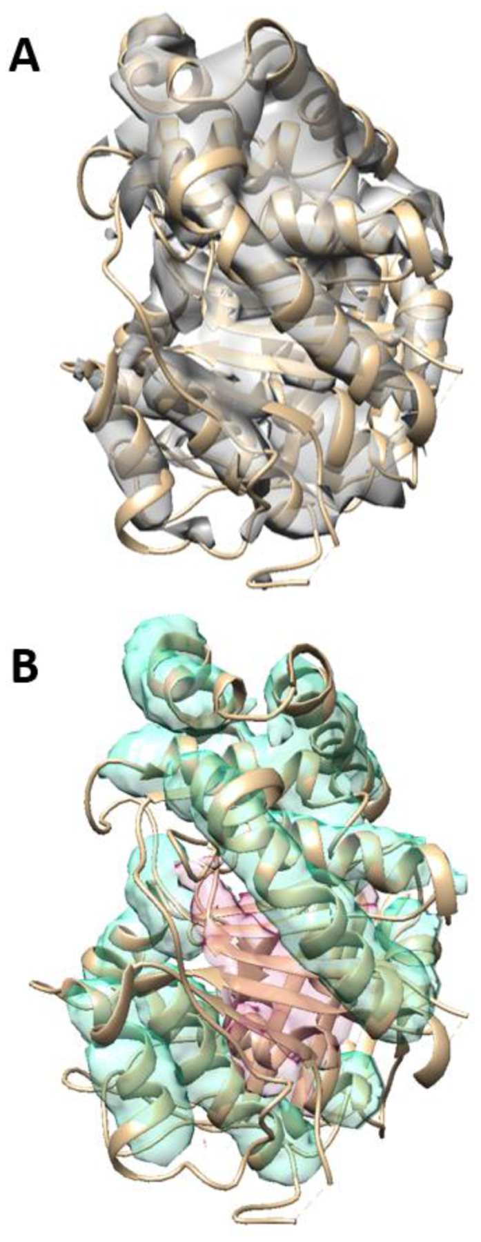
Figure 3. An example of secondary structure segmentation using the CNN architecture.

(A) A 3D image simulated using the atomic structure of protein 3j7i_a (PDB ID) (shown in ribbon). (B) The detected helix regions (cyan), and β-sheet regions (pink) are superimposed with the atomic structure (ribbon).
