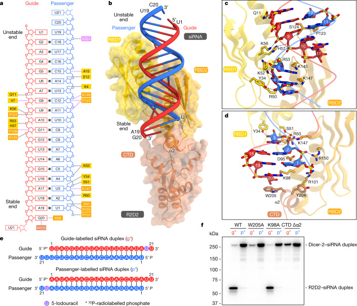
Fig. 4. siRNA recognition by R2D2.
a, Schematic of siRNA recognition by R2D2. The disordered nucleotides are depicted as dashed lines. b, Structure of R2D2 bound to siRNA. c,d, Recognition of the central region (c) and the stable end (d) of siRNA by R2D2. e, Nucleotide sequences of siRNA duplexes used in the photocrosslinking experiments in f. f, Photocrosslinking experiments. Dicer-2 and R2D2 (WT or mutants) were incubated with 5′-radiolabelled siRNA (g* or p*) bearing 5-iodouracil at position 20. The reaction mixture was analysed by SDS–PAGE, and crosslinked proteins were detected using phosphorimaging. CTD Δα2, the R2D2 mutant in which F201 and H215 are connected by a GGGS linker. n = 3 independent experiments.

