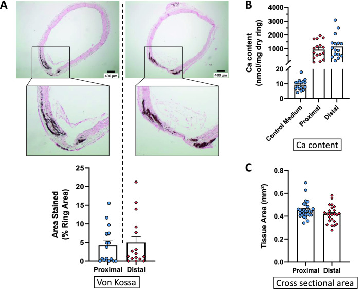
Figure 1.
Ex vivo calcification of rings isolated from the proximal (upper) and distal (lower) part of the descending thoracic aorta. (A) Representative examples of von Kossa staining of rings, individual values and mean ± SEM of stained areas expressed as percentage of the media area, after 14 days of culture in a calcifying medium containing high phosphate (3.8 mmol L−1) and 15% foetal calf serum. Scale bar = 400 µm, original magnification × 40. (B) Calcium content of the rings (mean ± SEM) cultured in calcifying medium or in control medium. (C) Cross sectional area of the rings (mean ± SEM). No significant differences were detected between both parts of the thoracic aorta.

