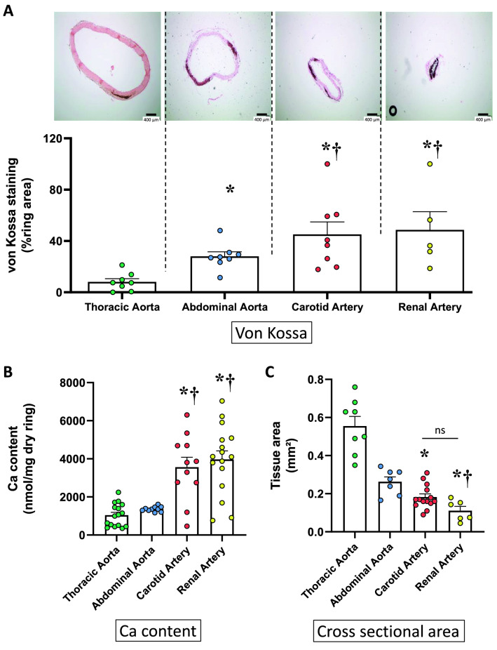
Figure 3.
Ex vivo calcification of rings isolated from various vascular beds. (A) Example of von Kossa staining of rings (without injury) and individual values and mean ± SEM of area stained after 14 days of culture in a calcifying medium containing high phosphate (3.8 mmol L−1) and 15% foetal calf serum. Scale bar = 400 µm, original magnification × 40. (B) Calcium content of the rings (mean ± SEM). (C) Cross sectional area of the rings (mean ± SEM). *p < 0.05 compared to thoracic aorta; †p < 0.05 compared to abdominal aorta. No difference was found between carotid and renal arteries; ns non-significant.

