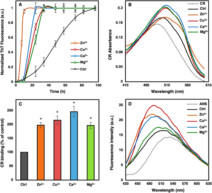
Figure 1.
α-Syn fibril formation in the presence and absence of each cation. 100 µM α-Syn was incubated under constant shaking (1000 rpm) in 20 mM Tris buffer (pH 7.5) supplemented with 500 µM of the mentioned metal ions at 37 °C. As a control (Ctrl), α-Syn was incubated in Tris buffer alone. (A) For kinetic studies, fibril formation was monitored by 25 µM ThT fluorescence excited at 450 nm. (B) Congo Red binding absorbance spectra of α-Syn fibrils formed in the presence and absence of the metal ions. (C) The amount of fibrils formed compared to control calculated based on bound CR using Eq. (2). (D) ANS fluorescence measurements were performed on end stage fibrils of each sample. Error bars present the standard deviation of three independent experiments. CR: Congo red solution. ANS: ANS solution.

