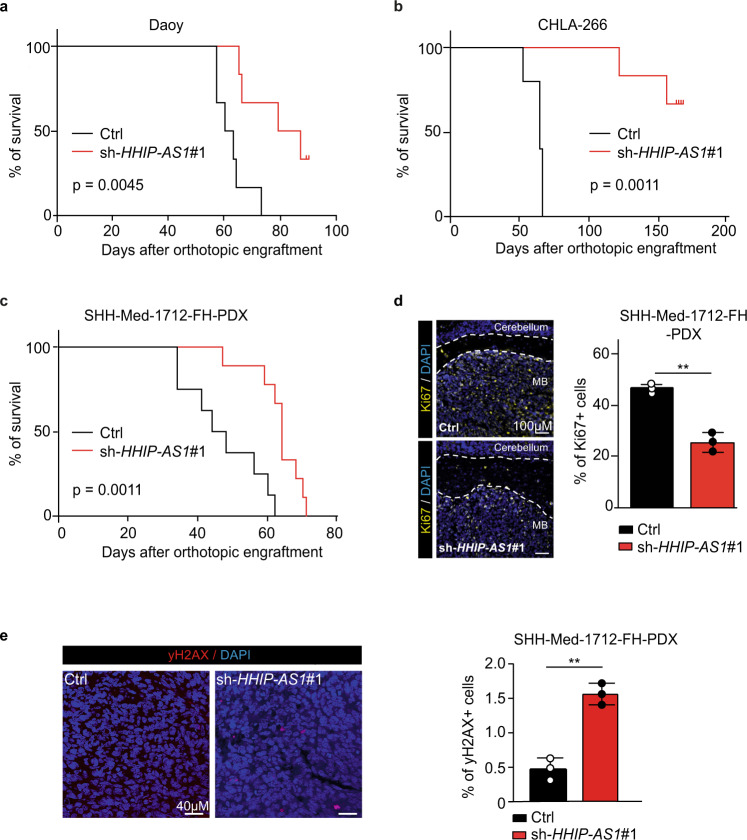
Fig. 6. Loss of HHIP-AS1 extends survival in SHH-driven brain tumor models in vivo.
a,b Kaplan–Meier estimates of nude mice orthotopically engrafted with stably HHIP-AS1-silenced (sh-HHIP-AS1#1) Daoy (a, n = 13 mice) and CHLA-266 (b, n = 13 mice) cells compared to corresponding controls (sh-scr), respectively. c Survival curves of nude mice orthotopically engrafted with transiently HHIP-AS1-silenced (sh-HHIP-AS1#1) SHH-Med-1712-FH cells (n = 17mice). For panels a, b, and c, the median survivals were compared using Mantel–Cox test. d,e Three controls and three HHIP-AS1-depleted SHH-Med-1712-FH tumors were immunostained for Ki67 (yellow, d, white scale bar: 100 µm), γH2AX (red, e, white scale bar: 40 µm) and the percentage of positive stained tumor cells is plotted in the bar graphs as mean ± SD. Nuclei are stained with DAPI (blue). Both, control (ctrl = sh-scr) and sh-HHIP-AS1#1 tumor tissue, are shown in the two representative images. Student’s two-sided t-test, **p < 0.01. Source data and exact p-values are provided as a “Source Data file”.

