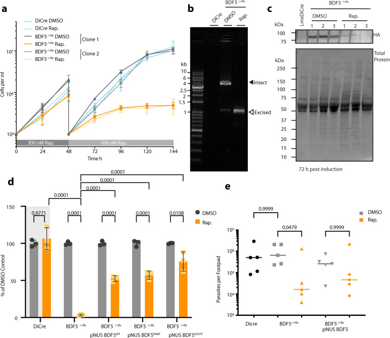
Fig. 2. Characterisation of inducible knockout of BDF5 using DiCre.
a Growth curve of promastigotes treated with the inducing agent, rapamycin (Rap.), or the vehicle, DMSO. For the first 48 h 300 nM rapamycin was added. At 48 h the cultures were passaged, and the concentration of rapamycin was lowered to 100 nM. Daily counting was conducted of triplicate cultures, of two independent clones, using a haemocytometer. Points and error bars denote mean values ± standard deviation, n = 3. b PCR and agarose gel analysis of BDF5::6xHAflx gene excision at the 72 h timepoint in (a). Solid arrowhead denotes the intact BDF5::6xHAflx gene and open arrowhead denotes the excised locus after rapamycin addition. The DiCre lane indicates the lack of PCR product in the parental strain. c Western blot showing levels of BDF5::6xHA protein after 72 h of DMSO or rapamycin treatment, conducted in biological triplicate. d Results of clonogenic survival assay comparing BDF5-depleted cells with cell lines carrying episomal complementation of BDF5 or mutated BDF5 alleles. Bars denote the mean of the percentage clonal survival where each experiment was normalised to its own DMSO control. Error bars indicate standard deviation, values above are p values from 2-way ANOVA with multiple comparisons by Tukey’s test, N = 3 replicate experiments. Lines denote comparisons performed by two-way ANOVA with associated p-values shown above. e Parasite burdens from infected mouse footpads determined by limiting dilution, individual points for each mouse with median values indicated by lines. Late-log cultures were pre-treated with 300 nM rapamycin and allowed to become stationary, prior to footpad infection for 8 weeks. Comparisons of Kruskal–Wallace test with Dunn’s correction indicated with associated p-values written above, n = 5.

