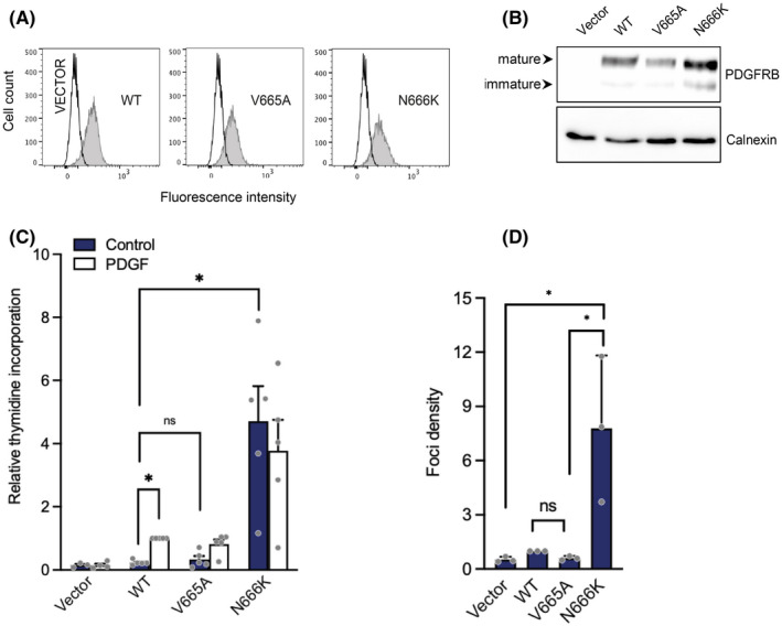
FIGURE 2.

p.V665A mutant receptor does not stimulate cell proliferation. (A) Ba/F3 cells were electroporated with PDGFRB (WT, V665A or N666K) or an empty vector. Cells were selected in the presence of G418 and sorted. The receptor cell surface expression was assessed by flow cytometry with a human PDGFRB‐specific antibody. (B) Transfected Ba/F3 cell lysates were analysed by Western blotting using anti‐PDGFRB antibodies. Membranes were re‐probed with an anti‐calnexin antibody as loading control. (C) Transfected Ba/F3 cells were stimulated with control medium, PDGF‐BB or IL‐3. After 20 h, [3H]‐thymidine was added to each well for 4 h. Incorporation of radiolabelled thymidine into cell DNA was quantified. Results were normalized using the stimulated wild‐type condition as reference. The average of five independent experiments is shown with SEM. All cell lines responded similarly to IL‐3 (not shown). (D) NIH3T3 cells were transfected in triplicates with PDGFRB (WT, V665A or N666K). Three weeks after transfection, foci were stained with crystal violet and quantified. The mean of three independent experiments is shown with SEM
