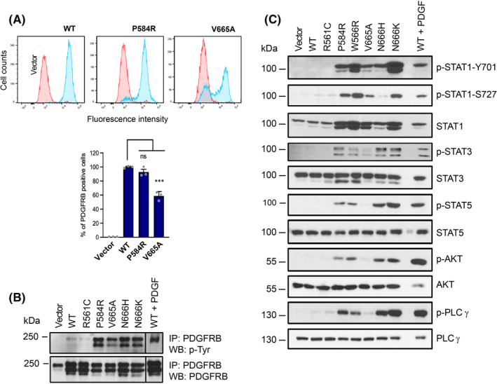
FIGURE 3.

p.V665A mutant preferentially activates STAT1. (A) Human PDGF receptor β expression in transduced NIH3T3 cells was tested by flow cytometry (blue) and compared to cells transduced with the empty vector (red). The average percentage of positive cells was calculated from three experiments with SEM. The receptor expression levels of the other cell lines used in the study are shown in Figure S1. (B) PDGF receptor β phosphorylation and expression levels. Transduced NIH3T3 cells were starved for 7 h. As a positive control, cells expressing WT receptors were stimulated with PDGF‐BB (25 ng/ml) for 15 min before lysis. The receptor was isolated by immunoprecipitation and analysed by Western blot using anti‐phospho‐tyrosine and anti‐PDGFRB antibodies. One representative blot out of three is shown. (C) NIH3T3 cells were treated as in (B). Total cell lysates were analysed by Western blot with the indicated antibodies (n = 3)
