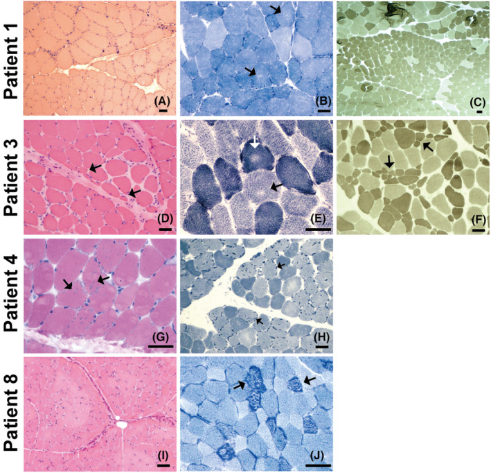
FIGURE 1.

Myopathological changes in CMS patients. Patient 1’s biopsy was taken from the left gastrocnemius muscles, and the others were taken from the left biceps brachii muscles. (A) HE staining showing increased fibre size variation and a group of severely atrophic angulated fibres. (B) NADH staining showing multiple and tiny areas of uneven oxidative staining and increased subsarcolemmal activity. (C) ATPase staining (pH 11.0) showing one fascicle composed of type 2 fibres with apparently reduced diameter. (D) HE staining showing increased fibre size variation with perifascicular fibre atrophy and increased endomysial fibrosis. (E) NADH staining showing uneven areas of oxidative reaction in both type 1 and type 2 fibres. (F) APTase staining (pH 11.0) showing type 2 fibre atrophy. (G) HE staining showing the presence of multiple vacuoles with a rim of basophilic material. (H) NADH staining showing multiple hyperintense dotty areas in type 2 fibres corresponding to tubular aggregates. (I) HE staining showing the slightly increased fibre size variation. (J) NADH staining showing multiple hyperintense dotty areas possibly corresponding to tubular aggregates. Scale bar = 50 μm
