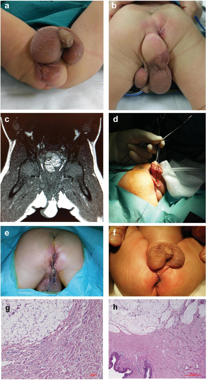
FIGURE 1.
Case 1. (a,b) Soft and spherical mass under the right hemiscrotum, with rugated pigmented scrotal skin on its upper part associated with incomplete penoscrotal transposition and right penoscrotal fusion. (c) Abdominal MRI: exophytic adipose tissue mass. (d) Surgery: excision of the mass and (e) perineal closure through interrupted resorbable stitches. (f) One-month follow-up. (g,h) Histological examination of specimens showing an area characterized by smooth muscle bundles dispersed in dermal collagen and a contiguous area with an abundant mature adipose tissue in the deep dermis and hypodermis.

