FIGURE 1.

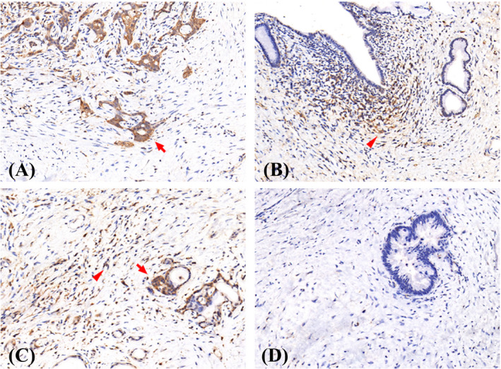
Representative microphotographs of CD9 staining in tumor (arrow) and stroma (arrowhead) area. (A) tumor+stroma‐, (B) tumor‐stroma+, (C) tumor+stroma+, (D) tumor‐stroma‐

Representative microphotographs of CD9 staining in tumor (arrow) and stroma (arrowhead) area. (A) tumor+stroma‐, (B) tumor‐stroma+, (C) tumor+stroma+, (D) tumor‐stroma‐