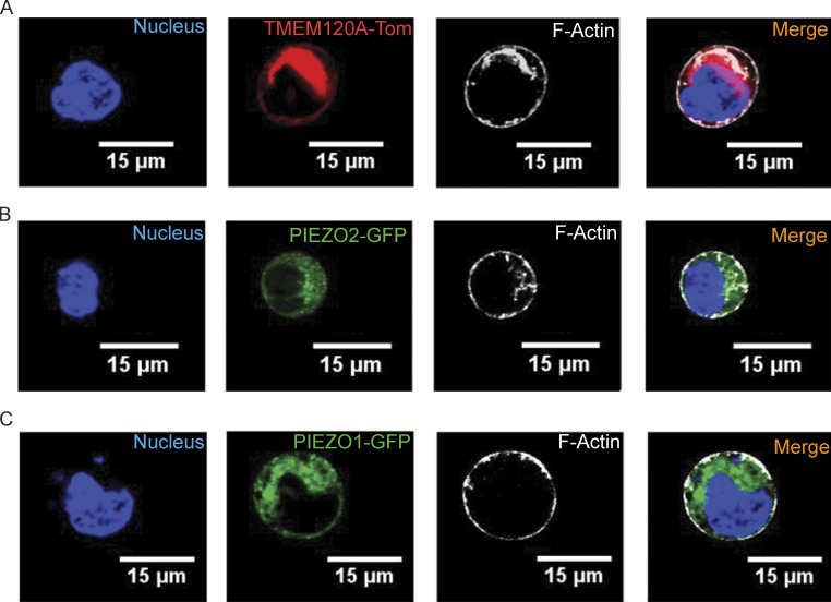
Figure S4.
TMEM120A is broadly distributed throughout cells. HEK293 cells were transfected with tdTomato-Tmem120a, GFP-Piezo1 or GFP-Piezo2, labeled with Sir-Actin, and confocal images were obtained as described in the Materials and methods section. (A) Representative confocal images of tdTomato-TMEM120A and Sir-Actin. (B) Representative confocal images of GFP-PIEZO2 and Sir-Actin, which labels F-Actin. (C) Representative confocal images of GFP-PIEZO1 and Sir-Actin. Two independent transfections were performed and 26–30 cells per group were imaged.

