Figure 4.
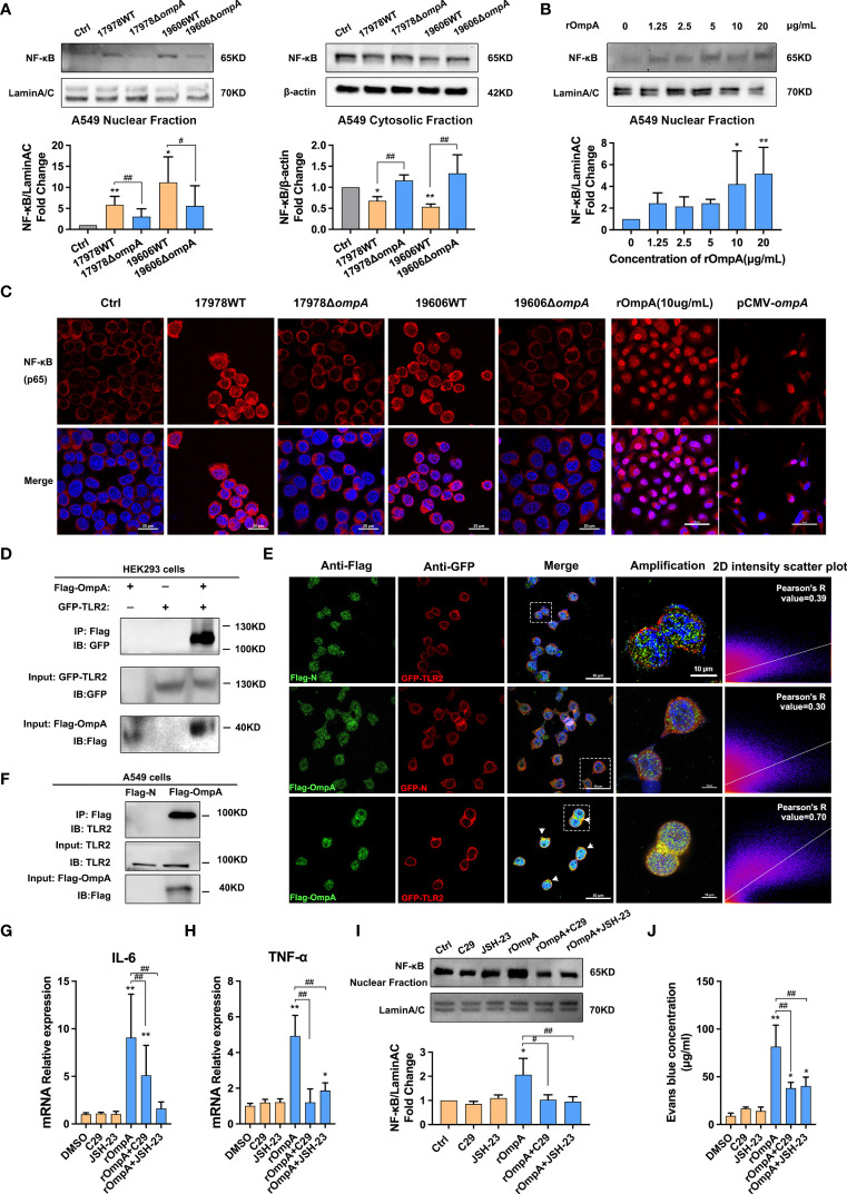
OmpA contributes to A. baumannii-induced TLR2/NF-κB activation for increased epithelial permeability. (A) Distributions of NF-κB in the cytoplasm and nucleus were evaluated by Western blotting. Relative NF-κB protein levels in the nuclei and cytoplasm are expressed relative to Lamin A/C and β-actin, respectively. (B) Distributions of NF-κB in the nuclei of A549 cells treated with different concentrations of purified OmpA (rOmpA). (C) The effects of different A. baumannii strains, rOmpA, and pCMV-ompA plasmid transfection on localisation of NF-κB were evaluated by immunofluorescence labeling, Red represents NF-κB and blue indicates nuclei. Scale bar: 20 μm. (D) Binding of OmpA-Flag and TLR2-GFP in HEK293 cells. (E) Co-localisation of OmpA-Flag and TLR2-GFP proteins was detected by double immunofluorescence staining in HEK293 cells, green represents Flag/OmpA-Flag, red represents GFP/TLR2-GFP, yellow represents OmpA-Flag/TLR2-GFP co-localization and blue indicates nuclei, scale bar: 50μm. Amplified images are shown below, scale bar: 10μm. (F) OmpA-Flag and TLR2 in A549 cells were detected by co-immunoprecipitation assay. (G–J) The effects of the TLR2 inhibitor, C29, and NF-κB inhibitor, JSH-23, on mRNA levels of IL-6 (G) and TNF-α (H), distribution of NF-κB in the nucleus (I), and EBA leakage (J) in A549 cells treated with rOmpA. **P<0.01,*P<0.05 vs. Control (or blank, DMSO) group, ## P<0.01, # P<0.05.

