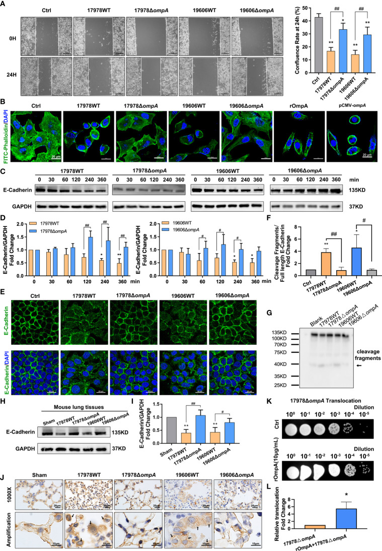
Figure 5.
OmpA induces cytoskeleton rearrangement and AJ internalisation for increased epithelial permeability and A. baumannii translocation. (A) Quantification of confluence rate of A549 cells at 24 hours after infection which represent the migration ability [% wound confluence = (a − b) × 100%/a; a = Initial scratch wound area at 0 h, b = Scratch wound area at 24 h], scale bar, 200μm. (B) FITC-phalloidin staining showed the cytoskeletal changes in A549 cells infected with different A. baumannii strains, treated with purified OmpA, or transfected with pCMV-ompA expression plasmid. Green represents phalloidin, and blue indicates nuclei. Scale bar: 20μm. (C, D) Expression of the adherens junction protein, E-cadherin, was evaluated by Western blotting. E-cadherin protein levels are expressed relative to GAPDH. (E) Localisation of E-cadherin was evaluated by immunofluorescence labeling. Green represents E‐cadherin and blue indicates nuclei. Scale bar: 20μm. (F, G) E-cadherin cleavage fragments (arrows) detected by the western blot, cleavage fragments levels are expressed relative to full length protein (135KD). (H–J) The expression level (H, I) and cellular localisation (J) of E-cadherin in the lung tissue of mice challenged by intratracheal injection of different A baumannii strains. (n = 3 per group), scale bar, 20μm (1000X), 10μm (Amplification). (K, L) Translocation of ΔompA strains across the A549 cell monolayer pre-treated with rOmpA protein. **P<0.01,*P<0.05 vs. Control (or Sham, blank, 17978△ompA) group, ## P<0.01, # P<0.05.

