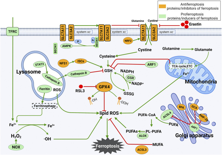
FIGURE 1.
Mechanism underlying the occurrence and regulation of ferroptosis. (1) Ferroptosis is mainly caused by lipid peroxidation. ROS leading to ferroptosis are produced by the iron-dependent Fenton reaction, mitochondrial electron transport chain or NOX proteins. Ferroptosis can be triggered by enhancing the synthesis of lipid ROS. (2) Inhibition of SLC7A11 deprives cells of cysteine, resulting in the loss of GSH and inactivation of GPX4. The latter further leads to the accumulation of lipid ROS and ferroptosis. The tricarboxylic acid cycle (TCA cycle) and electron carriers (ETC) in mitochondria stimulate GSH deficiency, thus leading to ferroptosis. The release of Fe2+ in mitochondria increases the level of free Fe2+ in cells and eventually promotes the production of lipid ROS. Lysosomal ROS contribute to the production of lipid ROS. In lysosomes, STAT3-mediated expression of cathepsin B is essential for ferroptosis via the MEK-ERK signaling pathway. In the Golgi apparatus, the Golgi stress response can inhibit ARF1, which is an inhibitor of GSH and ACSL4 and an activator of SLC7A11. Silencing ARF1 promotes ferroptosis by increasing cellular ROS levels.

