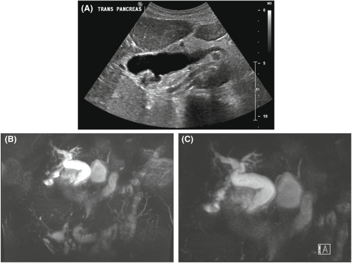
FIGURE 1.

Radiographic imaging of Case 1. (A) Transabdominal ultrasound from Case 1 demonstrating intrahepatic and extrahepatic biliary dilatation, multiple gallstones, and focal, irregular wall thickening without pericholecystic fluid. (B, C) Magnetic resonance cholangiopancreatography from Case 1 that demonstrates a prominent gallbladder and diffuse intrahepatic and extrahepatic biliary ductal dilatation
