
FIGURE 5.
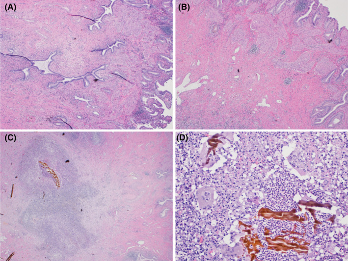
Microscopic imaging of Case 2. H&E‐stained sections demonstrating adenomyomatosis of the gallbladder with benign glands and associated smooth muscle bundle at 2× magnification (A) and 4× magnification (B). Also seen within the same specimen, an intramural abscess at 4× magnification (C) and associated xanthogranulomatous inflammation with foamy histiocytes and multinucleated giant cells surrounding inspissated bile (D) shown at 20× magnification