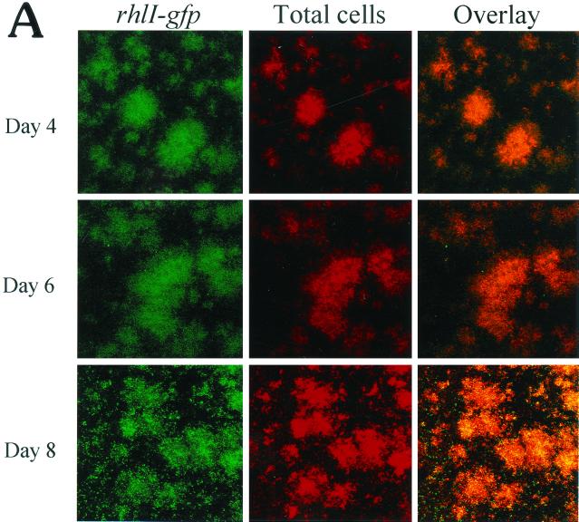
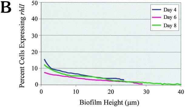
FIG. 4.
(A) Scanning confocal micrographs of biofilms formed by PAO1(rhlI-LVAgfp) grown for 8 days in a flowthrough chamber. Biofilms were examined on days 4, 6, and 8 to determine the number of cells expressing rhlI-LVAgfp relative to the total number of cells. Magnification, ×360. (B) Percentage of cells expressing rhlI plotted as a function of biofilm height.


