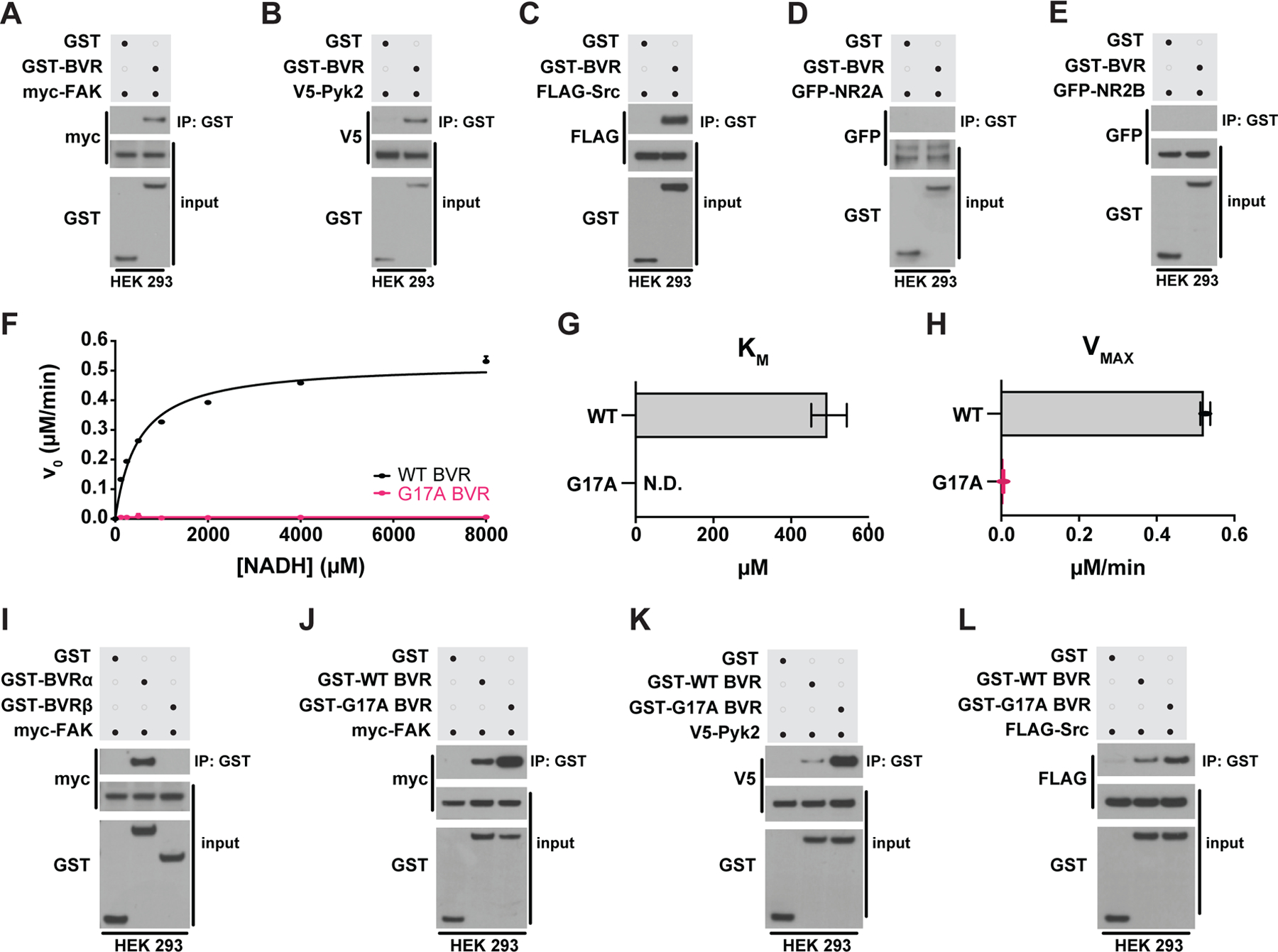
Figure 6. BVR physically interacts with FAK/Pyk2 and Src in a reductase-independent manner.

(A to E) Immunoblots of lysates (input) and GST immunoprecipitates (IP) from HEK 293 cells overexpressing GST or GST-BVR and either (A) myc-FAK, (B) V5-Pyk2, (C) FLAG-Src, (D) GFP-NR2A, or (E) GFP-NRF2B. (F to H) Michaelis-Mention kinetics of bilirubin production by WT and G17A mutant BVR with varying concentrations of NADH (F), with each enzyme’s calculated KM (G) and VMAX (H). (I) Immunoblots of lysates (input) and GST IP from HEK 293 cells overexpressing myc-FAK and either GST, GST-BVRα, or GST-BVRβ. (J to L) Immunoblots of lysates (input) and GST IP from HEK 293 cells overexpressing GST, GST-WT BVR, or GST-G17A BVR and either (J) myc-FAK, (K) V5-Pyk2, or (L) FLAG-Src. (Data are either representative of or quantified (mean ± SEM) from n = 3 independent experiments.
