Figure 4.
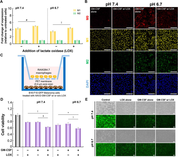
Effect of GM-CSF combined with LOX on macrophages or on cancer cell growth. (A) The fold changes in M1 and M2 marker expression relative to untreated macrophages (#p < 0.005, §p < 0.0005; two-tailed unpaired Student’s t test). The bars represent the mean ± standard deviation (n = 4). (B) Representative confocal images of macrophages treated as in (A). Scale bar = 100 μm. (C) Schematic representation of the coculture model established with AAV2-GM-CSF-infected B16-F10-GFP cancer cells (receiver well) and macrophages (membrane insert) for the measurement of cancer cell growth. (D) The cell viability of B16-F10-GFP cells cocultured with macrophages under various conditions using Transwell plates (*p < 0.05, §p < 0.0005, ‡p < 0.00005; two-tailed unpaired Student’s t test). The bars represent the mean ± standard deviation (n = 4). (E) Representative images of B16-F10-GFP cells cocultured with macrophages under various conditions in Transwell plates. Scale bar = 100 μm.

