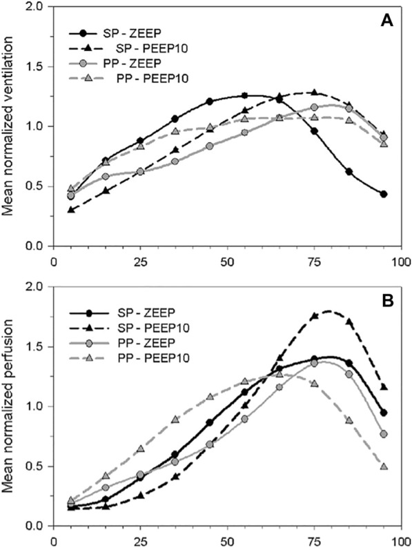
Fig. 4.

Prone position-induced changes, in interaction with PEEP, in ventilation and perfusion along the dorso-ventral gradient in an experimental model of ARDS. The figure shows the mean normalized values of regional ventilation (A) and perfusion (B), measured in PET by inhaled 13 N and injected [15O]-H2O, in 10 lung sections distributed along the dorso-ventral gradient of animals in the supine (SP) or the prone position (PP) and with zero end-expiratory pressure (ZEEP) or a PEEP of 10 cmH2O (PEEP) The figure shows that perfusion is redistributed to ventral lung regions, to a greater degree compared to regional ventilation, when a prone position is performed in conjunction with the application of PEEP. PEEP positive end-expiratory pressure, PET positron emission tomography, PP prone position, SP supine position, ZEEP zero end-expiratory pressure. Reproduced with permission from Wolters Kluwer Health [50]
