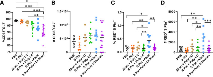
Figure 6.

B cell profile outside the germinal center after immunization. After three immunizations, lymphocytes from the draining popliteal lymph node macerated after intradermal immunization with S protein alone or with adjuvants [Poly(I:C); Alum; Poly(I:C) + Alum]. Controls were performed with PBS or Poly(I:C) + Alum. (A) Percentage of CD38+GL7- cells. (B) Number of CD38+GL7- cells. (C) Percentage of RBD+S Ptn+ cells. (D) Number of RBD+S Ptn+ cells. Figure representative of 4 independent experiments and shown as mean ± S.D. and was performed by one-way ANOVA with Tukey’s post hoc test. *p<0.05, **p<0.005, ***p<0.0005. (SEM; n=4-13).
