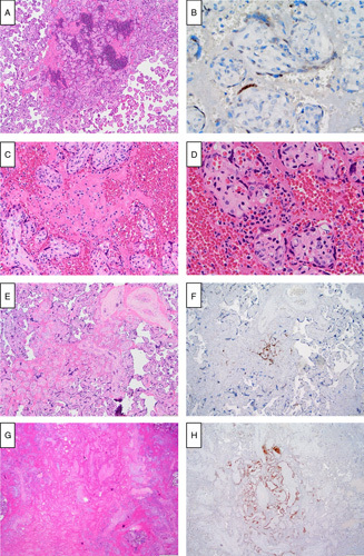
FIGURE 2.

Histologic features of early/localized SARS-CoV-2 placentitis with confirmation by IHC staining for SARS-CoV-2 spike protein. A, Localized, exudative pattern (case 5): sharply circumscribed focus of recent perivillous villous fibrin with scattered inflammatory cells and focal villous agglutination (hematoxylin and eosin). B, Localized, exudative pattern (case 5): cytoplasmic staining for SARS-CoV-2 capsid protein of a short, well-circumscribed strip of viable ScT (a few IHC-positive maternal leukocytes are seen in the intervillous space, other similar positive cells not shown) (SARS-CoV-2 immunostain). C, Localized, exudative pattern (case 5): maternal leukocytes (neutrophils, activated macrophages, and an eosinophil) embedded in a recent intervillous blood clot with focal ScT necrosis in adjacent villi (hematoxylin and eosin). D, Localized, exudative pattern (case 5): focally intense perivillous neutrophilic exudate with early ScT degeneration (hematoxylin and eosin). E, Localized, organizing pattern (case 6): small perivillous fibrin plaque with focally necrotic ScT (hematoxylin and eosin). F, Localized, organizing pattern (case 6): serpiginous strands of central positivity for SARS-CoV-2 capsid protein (SARS-CoV-2 immunostain). G, Localized, involuted pattern (case 7): ghost-like outlines of involuted villi surrounded by fibrinoid matrix and peripheral extravillous trophoblast (right). Inflammatory cells and viable ScT are not identified. (hematoxylin and eosin). H, Localized, involuted pattern (case 7): persisting positive staining for SARS-CoV-2 capsid protein outlines the contours of involuted central villi; surrounding villi, fibrinoid, and extravillous trophoblast lack staining (SARS-CoV-2 immunostain).
