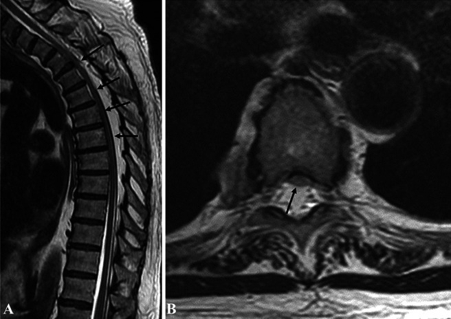
FIG. 1.

A: Sagittal T2-weighted MRI demonstrating epidural lipomatosis (black arrows pointing toward the cord compression by the lipomatosis) B: Axial T2-weighted MRI demonstrating anterior displacement and compression of spinal cord due to epidural lipomatosis (black arrow).
