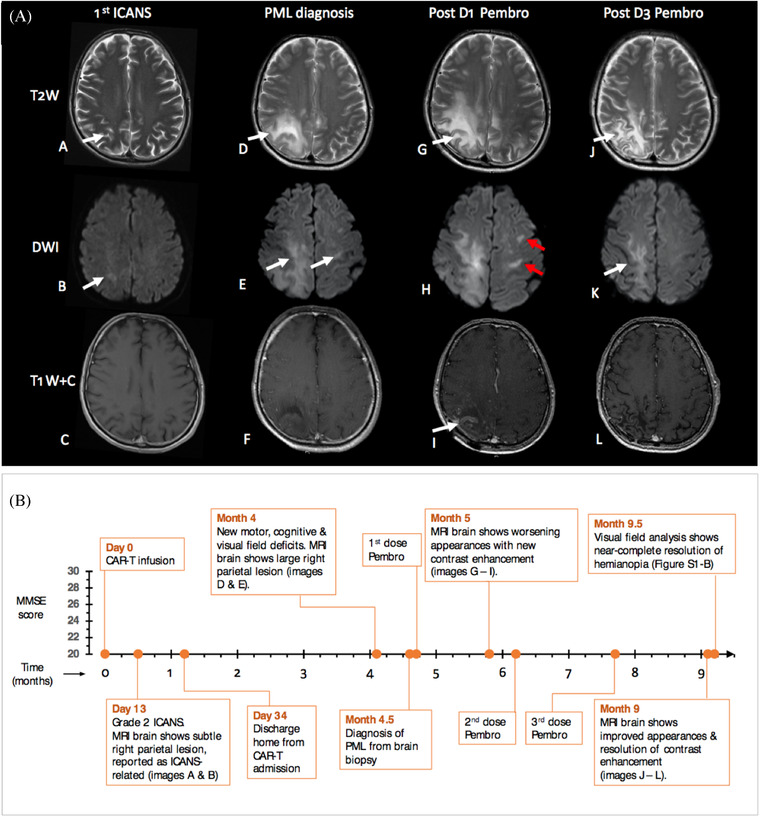
FIGURE 1.

(A) Magnetic resonance imaging of the brain. Axial, gadolinium‐enhanced images showing T2‐weighted (T2W), diffusion‐weighted (DWI) and contrast‐enhanced (T1W+C) sections at 1st episode of immune effector cell‐mediated neurological syndrome (ICANS; A–C), progressive multifocal leukoencephalopathy (PML) diagnosis (D–F), 1 month following 1st pembrolizumab dose (D1; G–I), and after 3rd dose (D3) of pembrolizumab (J–L), which was 5 months after 1st infusion. Panels A and B show a small focus of T2‐weighted signal abnormality in the right deep parietal white matter, demonstrating mild restriction of diffusion on DWI (B) at ICANS diagnosis. After the first pembrolizumab dose, note the relative increase in the size of PML lesions (G) with the progression of restricted diffusion (H) including new contralateral areas (red arrows) and new contrast enhancement (I), which had improved (J, K) or resolved (L) on interval imaging a month after D3.
(B) Timeline of clinical and radiological events post CAR‐T infusion (x‐axis) with serial mini‐mental state examination (MMSE) scores (y‐axis). At presentation, MMSE score was reduced (25/30), declined further after 2 weeks (22/30), but improved over 4 months (to 29/30) following pembrolizumab infusion. Image references refer to (A)
