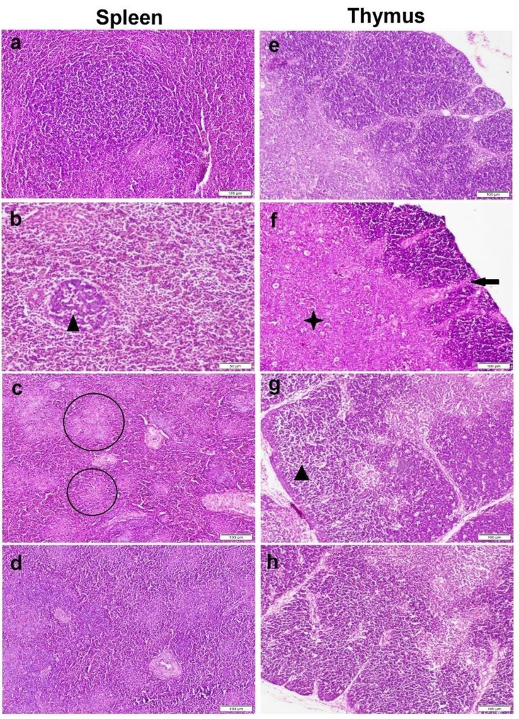
Fig. 6.

Photomicrograph of spleen (a–d) and thymus (e–h) tissue sections stained by haematoxylin and eosin. a and e – control group with normal histological structure; b and f, c and g – ochratoxin A (OTA) group showing lymphocytic cell depletion and lymphocytolysis (black triangles), multifocal areas of necrosis (circles), thinning in the cortical layer (black arrow) and expansion with extensive necrosis of medulla (black star); d and h – Bacillus subtilis fermentation extract + OTA group showing normal histological structure
