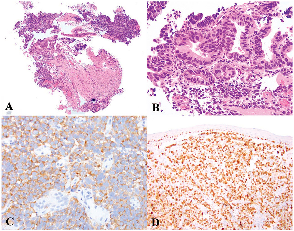
Figure 2.
Small cell neuroendocrine carcinoma (SCNEC). A and B, SCNEC of the esophagus demonstrating solid sheets of small cells with high N:C ratio, fusiform nuclei, nuclear molding and scant cytoplasm arising in a background of high-grade dysplasia in Barrett esophagus. C, Immunohistochemical staining for synaptophysin shows diffuse staining with focal “dot-like” positivity in some cells. D, Ki-67 immunostaining, with a Ki-67 proliferation index of 90% (hematoxylin-eosin, original magnifications ×2 [A] and ×100 [B]; original magnifications ×200 [C] and ×100 [D]).

