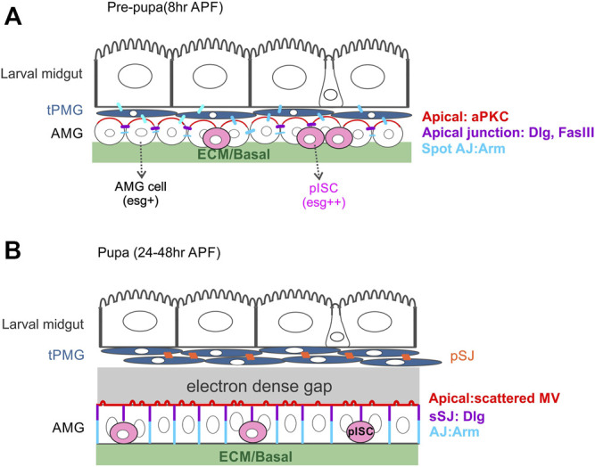
FIGURE 4.

The organisation of the midgut during pupal development. (A) During the first hours after puparium formation, the peripheral cells of late larval midgut AMP nests re-arrange to form the tPMG (dark blue) around the degenerating larval midgut cells. At the onset of metamorphosis, the central cells of late larval midgut AMP nests spread out to surround the tPMG. This layer of AMP cells initially express esg homogenously, but most AMG cells downregulate esg as they differentiate into ECs. A subset of AMPs maintain esg expression and become the presumptive intestinal stem cells (pISCs, pink), the precursors of the adult intestinal stem cells. At this stage, aPKC (red) localises to the apical domain of the AMG cells and Dlg and FasIII to the apical side of the lateral domain (purple). Spot AJs (blue) connect the larval ECs, the tPMG and the AMG cells. (B) From 20 h APF onwards, the tPMG appears as a tightly packed multi-layered structure with pleated SJs (orange) connecting the cells. By this stage, the tPMG has separated from the surrounding AMG and an electron dense liquid can be found between the two tissues. The AMG starts to develop irregularly spaced apical microvilli and smooth SJs at the apical side of the lateral membrane. AJs connect the more basal regions of the lateral membrane. pISCs remain basally localised.
