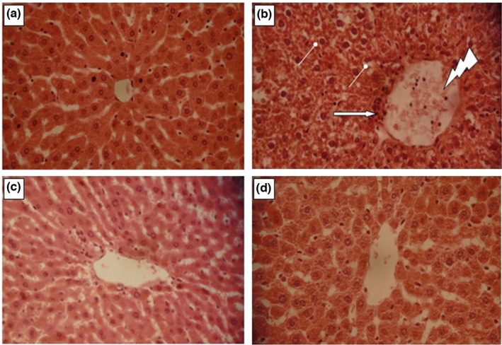
FIGURE 7.

Histopathological observation of liver tissues in both control and experimental animals (a). Group 1 served as control. (b) Group 2 rats were induced hepatic damage by daily intraperitoneal injection of CCl4 (1 ml/kg in 1% olive oil. i.p.) for 14 day. Arrow indicates leukocyte inflammatory cells. Congested central veins. Hepatocyte vacuolization. (c) Group 4 rats were pretreated daily with LmPS (250 mg/kg BW) for 14 days and then intoxicated with CCl4 on the 14th day (1 mg/kg BW CCl4). (d) Group 3 rats were daily received LmPS (250 mg/kg BW) for 14 days. Optic microscopy: HE (×400). Scale bars = 100 µm
