FIGURE 2:
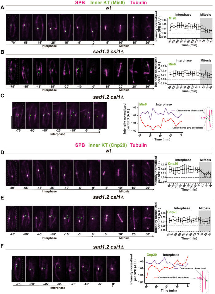
Mis6 and Cnp20 are stably associated with the centromeres in Rabl configuration-deficient cells. (A–F) Frames from films of mitotic cells carrying Sid4-mCherry (SPB), ectopically expressed mCherry-Atb2 (tubulin), and endogenously tagged Mis6-GFP, A–C, or Cnp20-GFP, D–F. Bars, 5 µm. Mean of total Mis6-GFP, A, B, and Cnp20-GFP, D, E intensities through interphase and mitosis were quantified. Ten cells during more than three independent experiments were monitored for focal intensity of Mis6-GFP and Cnp20-GFP. Error bars represent standard deviations; t = 0 min is just before SPB separation. (C, F) Quantification of Mis6-GFP and Cnp20-GFP focus intensity separately in centromeres with and without SPB association in sad1.2 csi1∆ cells. (A, D) In wt cells, all Mis6-GFP and Cnp20-GFP signals localize to the SPB during interphase. (B, C, E, F) sad1.2 csi1∆ cells showing centromere dissociation from the SPB also show stable signals through interphase.

