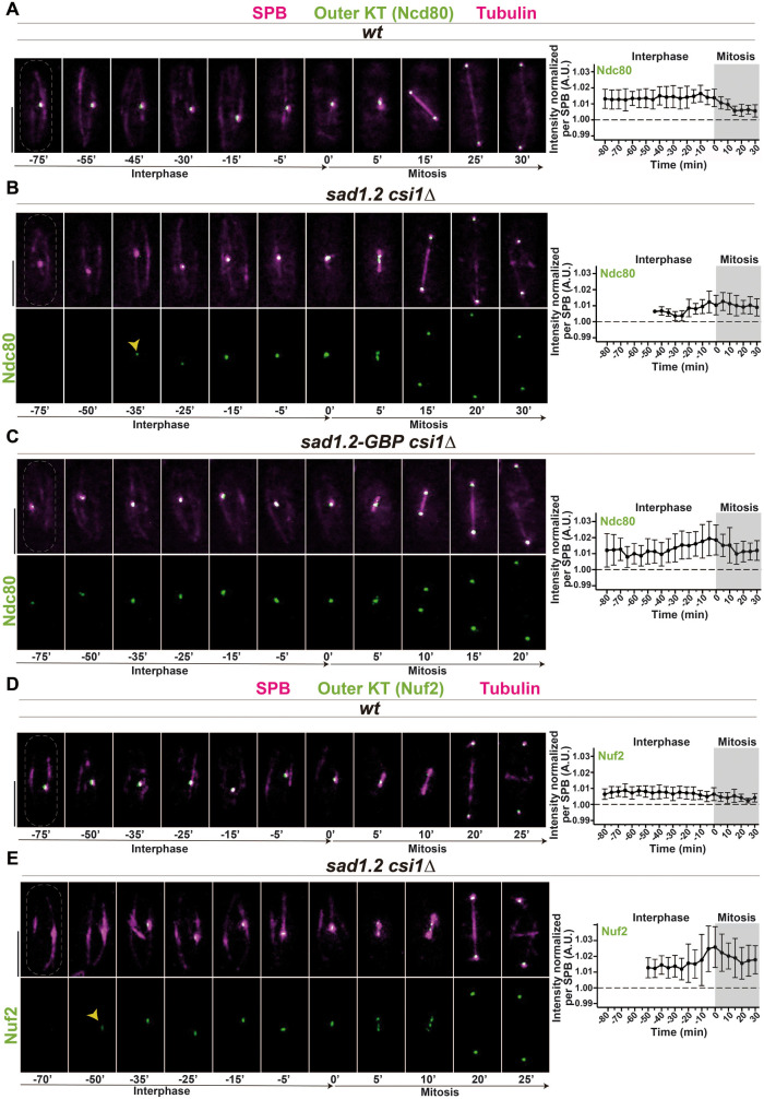
FIGURE 3:
Ndc80 and Nuf2 are dissociated from centromeres during interphase and reassembled at mitotic onset in sad1.2 csi1∆ cells. (A–E) Frames from films of mitotic cells carrying SPB and tubulin markers as in Figure 2, and endogenously tagged Ndc80-GFP, A–C, or Nuf2-GFP, D, E. Bars, 5 µm. Means of total Ndc80-GFP and Nuf2-GFP intensities were quantified as in Figure 2. Ten cells during more than three independent experiments were monitored. Error bars represent standard deviations; t = 0 min is just before SPB separation. (C) The GBP-GFP system was used to force centromere–SPB interactions (see Supplemental Figure 3C). Association with centromeres and levels of Ndc80 protein in interphase are recovered in sad1.2-GBP csi1∆ settings compared with wt settings.

