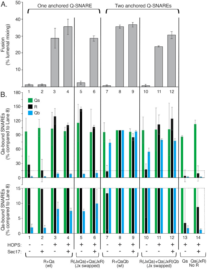
FIGURE 6:
The block to fusion from swapped Jx domains is relieved by Sec17 or by having wild-type Qb anchored to the membrane by its TM domain. Proteoliposomes with Ypt7 and SNAREs were prepared as described in Materials and Methods with R (wild-type) or R having the Jx region of Qa, with Qa (wild-type) or Qa having the Jx region of R, or with QaQb with wild-type Qa or with Qa having the Jx region of R. Fusion reactions and immunoprecipitations were conducted as described in Materials and Methods, with 100 nM Qc, 100 nM sQb (where Qb was not present on the proteoliposomes), 50 nM HOPS, and with or without 500 nM Sec17, as indicated. (A) Average FRETs with SDs are shown at 20 min for three replicates. (B) Samples from each incubation were solubilized in RIPA buffer and analyzed for Qa-bound Qb and R as described in Materials and Methods, and bands were analyzed with UN-SCAN-IT software for pixel intensity as compared with lane 8 with all wild-type SNAREs. Two y-axis scales are shown to display differences of bound sQb in lanes 1–6. Averages and SDs of the three replicates are shown. See Supplemental Figure S2 for typical kinetic data and a representative Western blot image.

