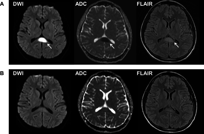
Fig. 1.
Magnetic resonance images of the brain of Patient 1 on Day 18 (A) and on Day 25 (B) after COVID-19 vaccination. A Diffusion-weighted image (DWI) (left) shows restricted diffusion in the splenium with low apparent diffusion coefficient (ADC) values (middle), and fluid-attenuated inversion recovery image (FLAIR) (right) shows a high signal intensity lesion at the midline of the splenium of the corpus callosum (arrows). B The lesion disappeared after intravenous high-dose methylprednisolone therapy

