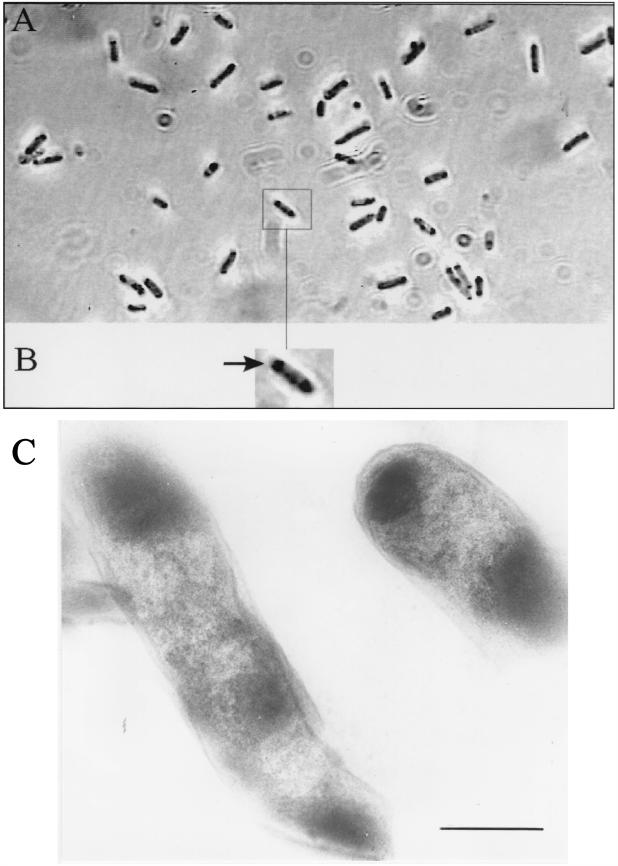
FIG. 1.
Light (A and B) (magnification, ×10,000) and electron (C) (magnification, ×43,000) microscopic images of recombinant E. coli(pSKBEC/PP:3.3). (A and B) E. coli cells were grown for 7 days on LB agar plates. Panel B shows an enlarged representative cell of E. coli; the arrow indicates the accumulated indigo. (C) Cells grown for 12 h in LB medium containing IPTG (1 mM), ampicillin (75 μg/ml), and tetracycline (12.5 μg/ml) and fixed as described previously. Bar = 0.5 μm.

