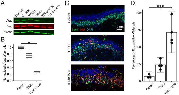
Fig. 2.
Proliferation of Müller glia in human retinal organoids after treatment with Lats kinase inhibitors. (A) An immunoblot for pYap and tYap after a 24 h incubation of human retinal organoids shows modest suppression of Yap phosphorylation after treatment with 10 μM TRULI and a profound effect of 3 μM TDI-011536 by comparison to controls containing DMSO, the solvation vehicle for the compounds. Each sample includes five organoids; β-actin serves as a loading control. (B) Quantification of the immunoblot by the ratio of pYap to tYap, both normalized to the DMSO control, confirms a significant effect of treatment with both compounds by one-way ANOVA; P = 0.019 for two experiments in each condition. (C) After 5 d of treatment, confocal immunofluorescence microscopy of sections from human retinal organoids reveals a substantial increase in cells doubly positive for EdU and Sox9, which represent proliferating Müller glia. (D) Quantification of the foregoing result demonstrates the significance of the effect by one-way ANOVA; P = 0.0003 for four experiments in each condition.

