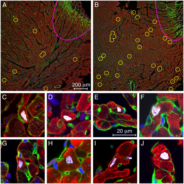
Fig. 5.
EdU incorporation into cardiomyocytes treated with TDI-011536. (A) At a distance from the lesion (pink arc at top right), a low-power micrograph of a representative section from a control animal shows only a few, small EdU-positive cells (circles). (B) After 3 d of treatment with TDI-011536, there are substantially more EdU-labeled cells outside the lesion, many of them large cardiomyocytes. (C–J) Individual EdU-labeled cardiomyocytes from treated animals are displayed. In all the images, cardiomyocytes are immunolabeled for troponin I or alpha-smooth muscle actin (red). Nuclei are stained with DAPI (blue) and those incorporating DNA precursors are additionally marked with EdU (white). By labeling membrane glycoproteins, wheat-germ agglutinin (green) helps to delineate the boundaries of cardiomyocytes. The small round profiles represent capillaries.

