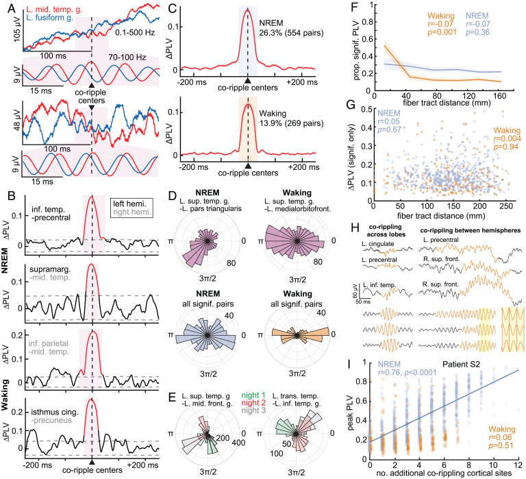
Fig. 4.
Ripples phase-lock across wide separations in the cortex. (A) Two example pairs of co-occurring ripples in broadband LFP and 70- to 100-Hz bandpass. A consistent phase lag from the left middle temporal gyrus (red) to left fusiform gyrus (blue) is evident in the expanded band-passed (70- to 100-Hz) 50-ms-long traces centered on the coripple (pink background). Similar phase lags were also present across other coripples between these sites, resulting in a significant PLV. (B) Example 70- to 100-Hz ΔPLV time courses calculated between ripples co-occurring between ipsilateral and contralateral cortical sites (≥25-ms overlap) in NREM and waking. Red shows significant modulation (post-FDR P < 0.05, randomization test, 200 shuffles per channel pair). (C) Average ΔPLVs (relative to −500 to −250 ms) for cortical channel pairs with significant PLV modulations. A greater percentage of pairs had significant coripple PLV modulations during NREM (26.3%) than waking (13.9%). (D) Polar histograms of waking and NREM 70- to 100-Hz phase lags across coripples for two example cortico-cortical pairs with significant PLV modulations (Top) and across channel pair circular means (Bottom). The magnitude of each pie wedge corresponds to the number of coripples (Top) or number of channel pairs (Bottom) with the indicated phase lag. cortico-cortical phase lags had a significant preference for ∼0 or ∼π during waking compared to NREM based on the counts within 0 ± π/6 or π ± π/6 vs. outside these ranges (P = 5 × 10⁻⁸, χ2 = 29.8, df = 1). (E) Example coripple phase-lag distributions for different sleep nights. The dominant phase lag for coripples changes across nights (color-coded). (F) Proportion of channel pairs with significant PLVs has a weak, nonsignificant decrease over distance during NREM but a significant decrease, notably at shorter distances, during waking (linear mixed effects with patient as random effect). See SI Appendix, Fig. S6 C and D for individual patients. (G) ΔPLV does not decrement with intervening fiber tract distance for channel pairs with significant coripple PLV modulations (linear mixed effects with patient as random effect). See SI Appendix, Fig. S6 E and F for results from individual patients. (H) Single-sweep broadband LFP and normalized 70- to 100-Hz bandpass show waking ripples (orange) co-occurring across lobes (Left) and between hemispheres (Right). Note the consistency of ∼0 or ∼π phases in the shaded inset. (I) Peak PLV correlates with the number of additional cortical sites corippling during NREM in a sample patient (n = 10/17 patients significant, post-FDR P < 0.05, significance of r) more often than waking (n = 3/17). Fit is linear least-squares regression. The PLVs between the indicated channels measure the consistency of phase between those channels at each latency relative to ripple peak across all instances of ripples co-occurring between those channels in NREM or waking.

