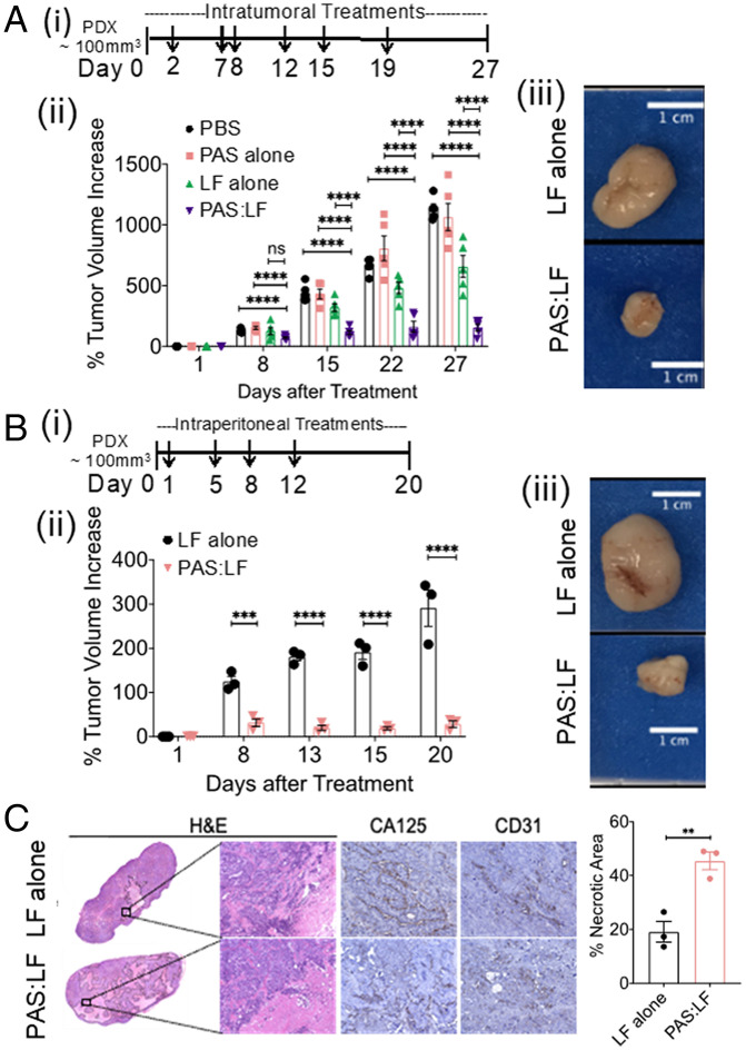
Fig. 7.
PAS:LF toxin reduces tumor burden in a PDX model of OvCa and is well tolerated in mice. The human ovarian PDX tumor (NCI:572874–153-T) was implanted and expanded in female NRG mice, then injected s.c. into three to five mice per treatment group. (A) Once tumors reached ∼100 mm3, (i) mice were treated with a total of 6 intratumoral injections (as indicated by arrows) of vehicle (PBS), 5 μg LF alone, 15 μg PAS alone, or 15 μg PAS:5 μg LF (5/group) over ∼3 wk. (ii) Tumor volumes were measured using calipers over time (****P < 0.001; ns, nonsignificant). Error bars represent SEM. (iii) Tumors were harvested at end point (representative images of harvested tumors are shown). (B) (i) PDX tumors were also treated with a total of four i.p. injections (indicated by arrows) of 5 μg LF alone or 15 μg PAS:5 μg LF (3/group) over 1.5 wk. (ii) Tumor volumes were measured using calipers over time (***P < 0.005, ****P < 0.001). Error bars represent SEM. (iii) Tumors were harvested at end point (representative images of harvested tumors are shown). (C) Harvested tumors treated with i.p. doses were paraffin embedded, sectioned, and H&E stained (representative images of 10X whole scan and 20X magnified area) (Left Panel). Serial sections were stained with CA125 and CD31. Percent necrotic area was quantified from H&E whole scans using ImageJ; data represent average values from three mice/group ± SEM (**P < 0.01 Right Panel).

