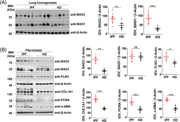
FIGURE 1.

BAG3‐mediated autophagy is insufficient in IPF fibroblasts. (A) Immunoblot analysis of BAG3, BAG1 or β‐actin from lung homogenates (LH) of IPF patients or healthy donors (HD) (left). The right part shows respective quantifications after normalizing their integrated density values (IDV) from n = 9 IPF patients and 5 HD. (B) Immunoblots from cell lysates of primary interstitial fibroblasts of IPF or HD for the indicated proteins. The right part shows respective IDVs that were normalized to β‐actin from n = 8 each for IPF and HD. p value summary: *p ≤0 .05, ** p ≤ 0.01, *** p ≤0.001.
Abbreviation: ns, not significant.
