
FIGURE 3.
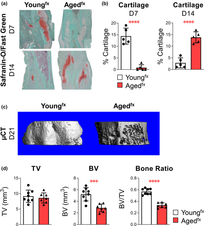
Bone fracture healing/tissue regeneration is delayed with advanced age. Young (4 months old) and aged (24 months old) mice underwent tibial fracture surgery. (a) Cartilage deposition was assessed by sectioning and staining 7‐ and 14‐day paraffin‐embedded fracture calluses with Safranin‐O/Fast Green. (b) The amount of cartilaginous tissue was determined using computer‐assisted histomorphometry. (c, d) Bone healing was assessed 21 days after injury using μCT analysis to measure TV, BV, and relative bone ratio (BV/TV). Cartilage analysis: youngfx, n = 5 and agedfx, n = 5 mice. Bone analysis: youngfx, n = 8 and agedfx, n = 7 mice. Data are expressed as mean ± standard deviation. Comparisons between youngfx and agedfx mice by an unpaired t test for each marker were performed with results indicated as ***p < 0.001 and ****p < 0.0001