FIGURE 6.

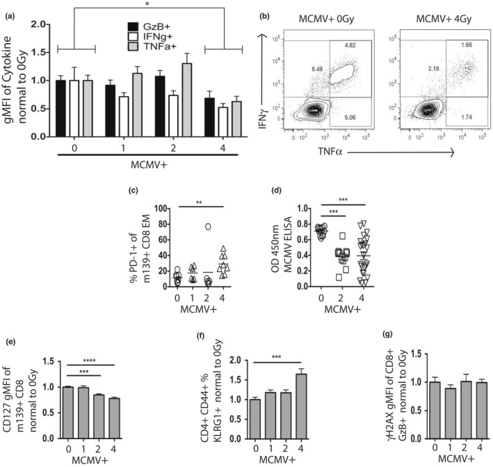
High‐dose sub‐lethal WBI in youth in MCMV+ mice taxes MCMV‐specific immunity. X‐axis: 0, 1, 2, and 4 = life‐long MCMV plus 0, 1, 2, and 4Gy in youth. (a‐b) Splenocytes from MCMV+ mice at 13 months of age were stimulated for 6 h with m139 peptide in the presence of brefeldin‐a. (a) Percent of individual cytokines normalized to 0Gy (no WBI) MCMV+ group. 2‐way ANOVA with Sidak's post‐test, 0Gy vs. 4Gy, p = 0.03. (B) Representative flow plots, as in (a). (c) Percent of PD‐1+ cells from PBMC at 13 months of age. Statistical outlier shown but not included in statistical analysis. (d) Results of MCMV ELISA from plasma collected at 19 months of age. (e) m139+ CD127 MFI of PBMC at 19 months of age from combined cohorts, normalized to 0Gy (no WBI) MCMV+ mice. (f) KLRG1‐hi percent of memory CD4 T cells from PBMC at 19 months of age from combined cohorts, normalized to 0Gy (no WBI) MCMV+ mice. (g) Gamma‐H2AX MFI of GzB+ CD8+ splenocytes at 13 months of age, normalized to 0Gy (no WBI) MCMV+ mice. (c‐g) Results of Dunnet's post‐tests shown
