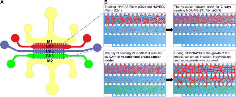
FIG. 1.
The design of the microfluidic chip and schematic diagram of the construction of the cancer model. (a) The materials were perfused in each channel as follows: Ch1: HUVECs + Fibrin gel; Ch2: HMCAF + Fibrin gel; Ch3: EGM (to form the self-assembled microvascular networks) or MDA-MB-231 + fibrin gel (to form a self-assembled vascularized breast cancer model); and M1 and M2: medium channels. (b) We obtained the functional self-assembled microvascular networks through this program and further constructed the vascularized breast cancer model.

