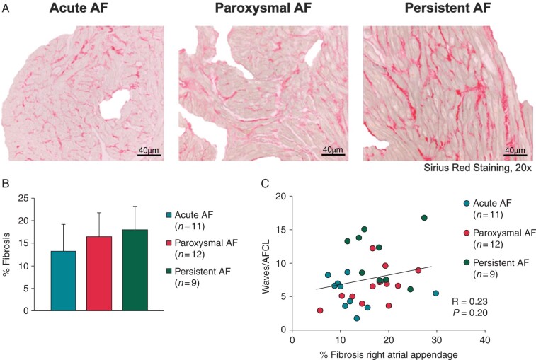
Figure 3.
(A) Representative pictures of Sirius red staining after exclusion of pericardial and perivascular fibrosis for aAF, pAF, and persAF. (B) Total amount of fibrosis (%) in the right atrial appendage. (C) Correlation between total amount of fibrosis (%) and AF complexity. AF, atrial fibrillation; aAF, acutely induced AF; pAF, paroxysmal AF; persAF, longstanding persistent AF.

