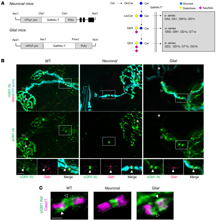
Figure 1. Anti–GM1 ganglioside antibody binding in transgenic mice with selective neuronal or glial complex ganglioside expression.
(A) Constructs used to direct GalNAc-T expression in neurons (human Thy1.2 promoter) or glia (mouse Plp promoter) of GalNAc-T–/–-Tg(neuronal) (Neuronal) and GalNAc-T–/–-Tg(glial) (Glial) mice, respectively. Ganglioside biosynthesis pathway indicates the reexpression of complex ganglioside expression (gray box) following construct insertion on a GalNAc-T–/– background (20). (B) Using a single anti-GM1 antibody (Ab, green), differential binding was observed at the distal motor nerves from triangularis sterni nerve–muscle preparations among genotypes. Open arrowheads indicate internodal Schwann cell (SC) abaxonal membrane anti-GM1 Ab deposition on WT and Glial nerves (absent along Neuronal nerves). Gliomedin (Gldn) immunostaining identifies the nodal gap. Boxed areas are enlarged underneath and represent differential anti-GM1 Ab binding at nodes of Ranvier (NoRs) among genotypes in relation to gliomedin (closed arrowheads). Dashed lines delineate the border of the axonal membrane determined by cytoplasmic CFP–positive axons. Scale bars: 10 μm (top panels) and 5 μm (lower panels). Asterisks indicate motor nerve terminals. (C) Caspr1 immunostaining (magenta) indicates the paranodes. Dashed lines delineate the border of the axonal membrane and arrowheads indicate anti-GM1 Ab binding beyond this membrane, suggesting binding on the glial membranes of the SC microvilli (open arrowheads) or paranodal loops (closed arrowheads) at WT and Glial NoRs. Scale bar: 2 μm.

