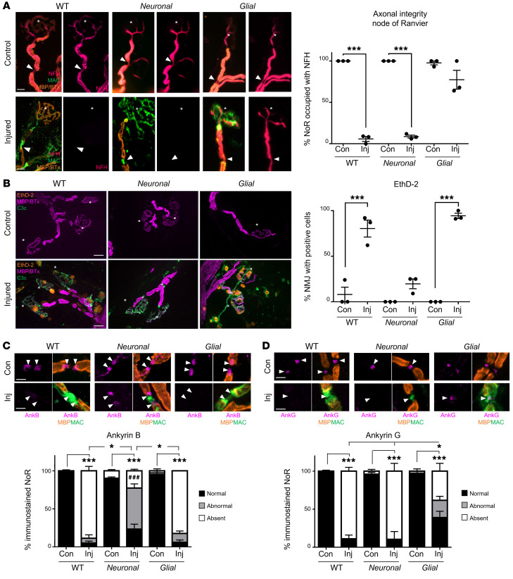
Figure 2. Distal motor nerve integrity following selective targeting and acute injury of neural membranes ex vivo.
Triangularis sterni nerve–muscle preparations from WT, Neuronal, and Glial mice were treated ex vivo with anti-GM1 Ab and a source of complement (injury, Inj) or anti-GM1 Ab alone (control, Con). (A) Loss of axonal integrity due to injury at the motor nerve terminal (MNT, identified by α-bungarotoxin, BTx, orange, asterisk) and node of Ranvier (NoR, orange, arrowheads) was monitored by presence of neurofilament H immunostaining (NFH, magenta). Membrane attack complex (MAC) complement pore deposition (green) was present in all injured preparations compared with control. (B) Ethidium homodimer–positive (EthD-2–positive, orange) cells overlying MNT (magenta, asterisk) were compared among treatment groups. Representative images show the presence of complement deposition (green) in all injured tissue. (C and D) The sites where ankyrin B (AnkB) or AnkG immunostaining should be located are indicated by arrowheads. The presence of normal (black bars, statistical comparisons indicated with asterisks) or abnormal (gray bars) AnkB and AnkG immunostaining was compared to associated controls for each genotype. A lengthened gap between AnkB domains is shown in a representative image from injured Neuronal tissue. Weakened, uneven AnkG staining in injured Glial tissue is shown in the representative image. Scale bars: 20 μm (A), 50 μm (B), and 5 μm (C and D). Results are represented as the mean ± SEM. n = 3/genotype/treatment: 10–46 NoRs/mouse (median = 21, NFH); 11–29 neuromuscular junctions (NMJs)/mouse (median = 18, EthD-2); 10–26 NoRs/mouse (median = 23, AnkG); and 12–31 NoRs/mouse (median = 21, AnkB) were analyzed. *P < 0.05; ***P < 0.001; ###P < 0.001 (for abnormal AnkB and AnkG immunostaining in C) compared with control by 2-way ANOVA with Tukey’s post hoc test.

