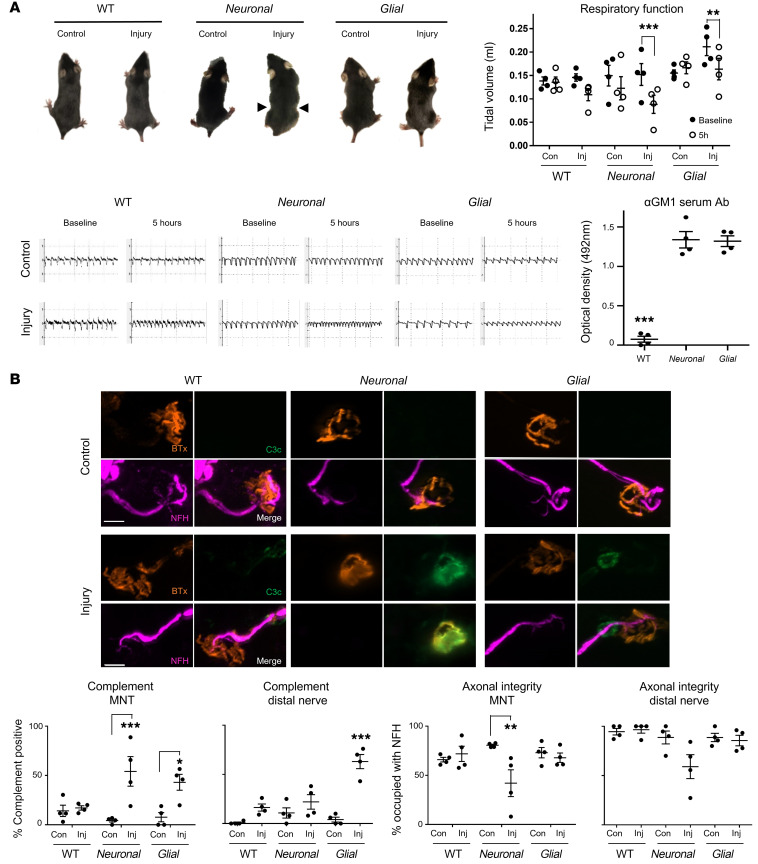
Figure 4. Distal motor nerve axonal integrity remains intact following selective glial membrane targeting in vivo.
WT, Neuronal, and Glial mice were dosed i.p. with 50 mg/kg anti-GM1 Ab followed 16 hours later with 30 μL/g normal human serum (NHS) (injury, Inj) or NHS only (control, Con). Respiratory function was monitored and diaphragm distal nerves assessed by immunoanalysis 5 hours after NHS delivery. (A) Injured Neuronal mice displayed the most severe respiratory phenotype: a pinched, wasp-like abdomen (arrowheads) and significantly reduced tidal volume (TV) measured using whole-body plethysmography (EMMS). Injured Glial mice also had significantly reduced TV compared with baseline. Representative respiratory flow charts for each treatment group show reduced TV and an increase in respiratory rate. Serum analysis indicates that circulating anti-GM1 Ab could be detected in Neuronal and Glial but not WT mice. Results are represented as the mean ± SEM, n = 4/genotype/treatment. (B) Complement deposition and axonal integrity (neurofilament H [NFH] occupancy) were compared at the diaphragm motor nerve terminals (MNTs) and along distal nerves. Representative images illustrate complement deposits (green) overlying the MNT, identified by bungarotoxin (BTx, orange), in injured Neuronal mice, and on the distal nerve in injured WT and Glial mice. Scale bar: 10 μm. Results are represented as the mean ± SEM. n = 4/genotype/treatment: 68–133 MNTs/mouse (median = 103) and 7–30 NoRs/mouse (median = 15) were analyzed. *P < 0.05, **P < 0.01, ***P < 0.001 by repeated measures 2-way ANOVA with Bonferroni post-hoc tests (A) or 2-way ANOVA with Tukey post-hoc tests (B).

